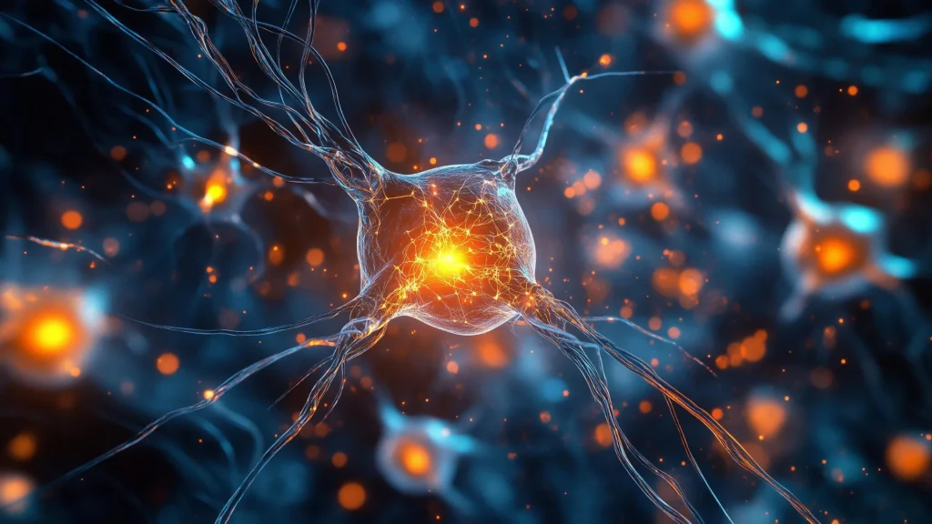
Scientists have developed a new imaging technique that reveals the hidden protein clusters driving Parkinson’s disease. This breakthrough could revolutionize early detection and uncover how the disease takes hold in the brain. Credit: Shutterstock
For the first time, researchers have directly seen and measured the protein clusters thought to spark Parkinson’s disease, marking a major milestone in understanding the world’s fastest-growing neurological condition.
These microscopic clusters, known as alpha-synuclein oligomers, have long been suspected as the starting point for Parkinson’s, but they have remained undetectable in human brain tissue — until now.
A team from the University of Cambridge, UCL, the Francis Crick Institute, and Polytechnique Montréal developed a powerful imaging approach that allows scientists to visualize, count, and compare these protein clumps in human brain tissue. One researcher described the breakthrough as “like being able to see stars in broad daylight.”